The Brain: What’s Going On in There? Lesson 1
Source: National Institute on Drug Abuse (1996) The Brain & the Actions of Cocaine, Opiates, and Marijuana. Slide Teaching Packet for Scientists
.Basic Science Health Connection
The brain controls virtually everything humans experience, including movement, sensing our environment, regulating our involuntary body processes such as breathing, as well as controlling our emotions. Ongoing scientific research into the organization and function of the brain has led, and will continue to lead, to new treatments of diseases such as Parkinson’s disease, epilepsy, stroke, and mental illnesses (including depression and schizophrenia).
The brain is the organ of behavior. It is also the organ of our mind. Both overt behavior and consciousness are manifestations of the work of our brains. Other people can see an individual’s overt behaviors, whereas consciousness is apparent only in our individual minds. The field of neuroscience studies how people control their behaviors, thoughts, and feelings, and how these actions some-times get out of control.
The brain processes a huge amount of information in a remarkably efficient manner. Think about driving a car. It is something most of us do without much difficulty. But to do it properly, we must perform a remarkable number of tasks. First we have to make sure that our body is in working order. Heart rate and breathing have to be properly regulated, body temperature held steady, and we certainly have to be sure we don’t fall asleep. Despite the complexity of these tasks, we carry them out with no conscious involvement on our part. Then, there are the things we are aware of. We have to see the road and hear the traffic (or the radio), use information from our feet, legs, hands, and arms to know where the gas pedal and steering wheel are, and then generate the body movements to control the direction and speed of the car. All of this often takes place while we are talking to someone else in the car, or even while talking on the phone (although this may not be the best idea). The magnitude and speed of data processing needed to do this are stunning, but most of us consider driving to be an easy task.
Different Regions of the Brain Regulate Different Functions
How does the brain carry out multiple tasks at one time? The answer is that the brain splits the larger task driving, in our example into smaller ones: seeing, hearing, moving, and so forth. Even those tasks are split into their component parts. One part of the human brain analyzes the movement of objects that we see, while another part is responsible for actually recognizing them. In short, specific parts of the brain carry out specific tasks. Not only that, but each part of the brain specializes in a specific kind of task. This means that whenever that task needs to be done, the appropriate information is processed by that part of the brain.
The flip side of this organizational scheme is that if a part of the brain is dam-aged, then the job it used to undertake cannot be done. For example, damage to the occipital lobe at the back of the brain can cause blindness, but it has no effect on a person’s ability to hear or move. Because the job of seeing is highly compartmentalized, individuals who have lost one aspect of sight, such as the ability to see colors or to recognize faces, may still be able to do other visual tasks. Imagine being able to recognize someone by hearing his or her voice, but not being able to recognize his or her face when you see it.
The advantage of this localization of function is when larger jobs are parceled out throughout the brain, they all can be done at once. This "division of labor" adds great speed to our ability to perceive what is happening in the world around us, to analyze it, and then to generate appropriate responses. Dealing with information in this way is called parallel processing. Computer scientists have used this concept in the development of computers.
The human brain consists of several large regions each of which is responsible for some of the activities necessary for life. These include the brainstem, cerebellum, limbic system, diencephalon, and cerebral cortex. The brainstem is the part of the brain that connects the brain and spinal cord.
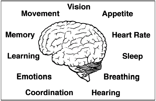
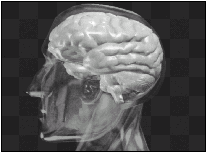
Figure 1.1: The human brain regulates everything a person does.
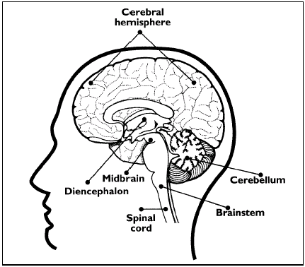
Figure 1.2: This drawing of a brain cut in half illustrates some of the major regions of the brain. Source: National Institute on Drug Abuse (1997) Mind Over Matter: The Brain’s Response to Drugs, Teacher’s Guide.
This part of the brain is involved in coordinating many basic functions such as heart rate, breathing, eating, and sleeping.
The cerebellum coordinates the brain’s instructions for skilled repetitive movements and for maintaining balance and posture.
The limbic system, as discussed in the next section, is involved in regulating emotions and motivations. In addition, parts of the limbic system, the amygdala and hippocampus, are important for memory functions.
The diencephalon contains the thalamus and hypothalamus. The thalamus is involved in sensory perception and the regulation of movement. The hypothalamus is an important regulator of the pituitary gland.
The cerebral cortex makes up the largest part of the brain mass and lies over and around most of the other brain structures. It is the part of the brain that is responsible for thinking, perceiving, and producing and understanding language. The cortex can be divided into areas that are involved in vision, hearing, touch, movement, smell, and thinking and reasoning.
Drugs Act On the Reward System in the Brain
Just as specific areas of the brain control seeing and hearing, specific brain areas also regulate emotions and motivations. These functions are carried out by a part of the brain called the limbic system. The limbic system, similar to other regions in the brain, influences how we respond to the world around us. Imagine a cool sunny day.

Figure 1.3: This drawing of a brain cut in half illustrates the lobes of the cerebral cortex and describes their main functions. Source: National Institute on Drug Abuse (1997) Mind Over Matter: The Brain’s Response to Drugs, Teacher’s Guide.
You finish your work early and head to your favorite park for a leisurely walk with your dog. You are feeling so mellow that you merely scratch the dog behind the ears when he slobbers on your clean shirt. You might have a very different reaction on another day when you have to work late, traffic is backed up, and the dog runs away instead of coming to welcome you home. This time when the dog slobbers on you (after he finds his way home again), you shove him away and scold him.
The feelings you have in those two different situations are a result of your limbic system at work. The limbic system uses memories, information about how your body is working, and current sensory input to generate your emotional responses to current situations.
The limbic system is involved in many of our emotions and motivations, particularly those related to survival, such as fear and anger. The limbic system also regulates feelings of pleasure related to our survival, such as those experienced from eating and sex. The feelings of pleasure, which scientists call reward, are very powerful. If something is pleasurable or rewarding, you want to do it again. Life-sustaining activities such as eating and sex activate a circuit of specialized nerve cells that are devoted to producing and regulating pleasure. These cells are located at the top of the brainstem in the ventral tegmental area (VTA). These neurons relay their messages through their axons to nerve cells in a limbic system structure called the nucleus accumbens. Additional nerve fibers reach part of the frontal region of the cerebral cortex. This circuit of neurons is called the reward system.
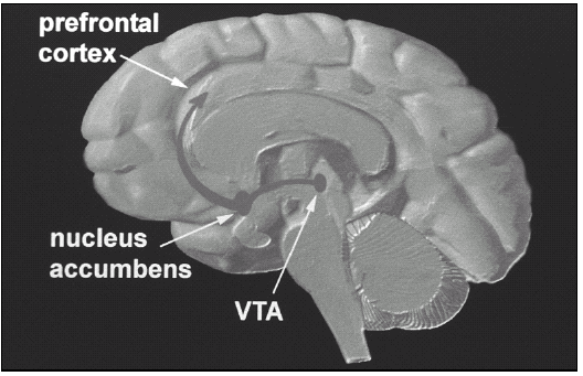
Figure 1.4: This drawing of a brain cut in half illustrates the brain areas and systems involved in the reward system, or pleasure circuit. Neurons in the ventral tegmental area (VTA) extend axons to the nucleus accumbens and part of the prefrontal cortex. Source: National Institute on Drug Abuse (1996) The Brain & the Actions of Cocaine, Opiates, and Marijuana. Slide Teaching Packet for Scientists.
Drugs of abuse activate these same VTA and nucleus accumbens neurons; that is why drugs produce pleasurable feelings to the drug user. And, because the feelings are pleasurable, the user wants to continue to experience the pleasure that he or she felt during previous drug use. One of the reasons that drugs of abuse can exert such powerful control over our behavior is that they act directly on the more evolutionarily primitive brainstem and limbic structures, which can override the cortex in controlling our behavior. Different drugs of abuse affect the neurons of the reward system in different ways. The activities in Lesson 3 in this module will elucidate the mechanisms by which drugs of abuse exert their effects. Imaging the Brain Scientists increasingly use newer technologies to learn more about how the brain works and how drugs of abuse change how the brain works. Historically, scientists could examine brains only after death, but new imaging procedures enable scientists to study the brain in living animal, including humans. One of the most extensively used techniques to study brain activity and the effects of drugs on the brain is positron emission tomography (PET). PET measures the spatial distribution and movement of radioisotopes in tissues of living subjects. Because the patient is awake, the technique can be used to investigate the relationship between behavioral and physiological effects and changes in brain activity. PET scans can detect nanomolar concentrations of tracer molecules and achieve spatial resolution of about 4 millimeters. In addition, computers can reconstruct images obtained from a PET scan in two or three dimensions. PET requires the use of compounds that are labeled with positron-emitting isotopes.4,5 A cyclotron accelerates protons into the nucleus of nitrogen, carbon, oxygen, or fluorine to generate these isotopes. The additional proton makes the isotope unstable. To become stable again, the proton must break down into a neutron and a positron. The unstable positron travels away from the site of generation and dissipates energy along the way. Eventually, the positron collides with an electron leading to the emission of two gamma rays at 180 degrees from one another. The gamma rays reach a pair of detectors that record the event. Because the detectors respond only to
simultaneous emissions, scientists can precisely map the location where the gamma rays
are very short-lived. The half-life (the time for half of the radioactive label to disintegrate) of the commonly used radioisotopes ranges from approximately two minutes to less than two hours, depending on the specific compound. Because a PET scan requires only small amounts (a few micrograms) of short-lived radioisotopes, negative pharmacological effects are imperceptible.
PET scans can answer a variety of questions about brain function, including the activity of neurons. Scientists use different radiolabeled compounds to investigate different biological questions. For example, radiolabeled glucose can identify parts of the brain that become more active in response to a specific stimulus. Active neurons metabolize more glucose than inactive neurons. Active neurons will emit more positrons. This will show as red or yellow on PET scans compared to blue or purple in areas where the neurons are not highly active. PET also helps scientists investigate how drugs affect the brain by enabling them to:
. determine the distribution of a drug in the body,
. measure the local concentration of a drug at binding sites,
. estimate receptor occupancy based on competitive binding assays,
. evaluate the effects of drugs on other neurotransmitter systems, and
. investigate the activity of enzymes that metabolize the drug.6
In addition to its uses in research, PET also is a powerful tool for diagnosing
Figure 1.5: When an unstable positron collides with an electron, the particles are annihilated and two gamma rays are emitted at 180° from each other. Detectors record gamma ray emission to localize the site of positron annihilation.
This material is based on work supported by the National Institutes of Health under Contract No: 263-98-C-0056. Any opinions, findings, conclusions, or recommendations expressed in this publication are those of the authors and do not necessarily reflect the view of the funding agency.
Copyright c 2000 by BSCS and Videodiscovery, Inc. All rights reserved. You have the permission of BSCS and Videodiscovery, Inc. to reproduce items in this module (including the software) for your classroom use. The copyright on this module, however, does not cover reproduction of these items for any other use. For permissions and other rights under this copyright, please contact BSCS, 5415 Mark Dabling Blvd., Colorado Springs, CO 80918-3842, www.bscs.org, info@bscs.org, (719) 531-5550.
NIH Publication No. 00-4871
ISBN: #1-929614-05-5