Neurons, Brain Chemistry,
and Neurotransmission
Lesson 2
Source: National Institute on Drug Abuse (1996) The Brain & the Actions of Cocaine, Opiates, and Marijuana. Slide Teaching Packet for Scientists.
The Brain Is Made Up of Nerve Cells and Glial Cells
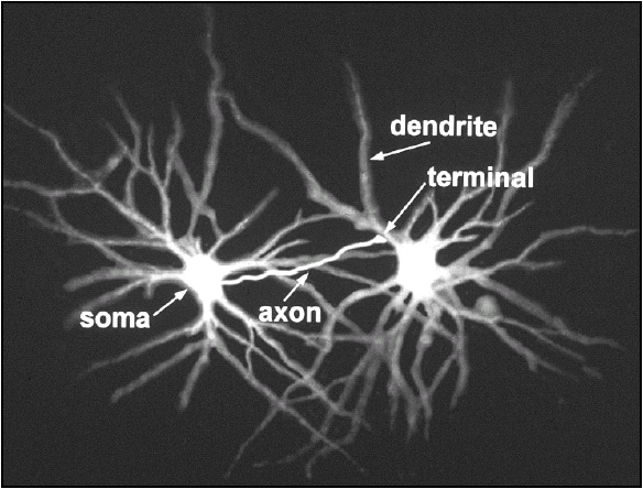
The cell body, also called the soma, is the metabolic center of the neuron. The nucleus is located in the cell body and most of the cell’s protein synthesis occurs in the cell body. A neuron usually has multiple processes, or fibers, called dendrites that extend from the cell body. These processes usually branch out somewhat like tree branches and serve as the main apparatus for receiving input into the neuron from other nerve cells. The cell body also gives rise to the axon. Axons can be very long processes; in some cases, they may be up to one meter in length. The axon is the part of the neuron that is specialized to carry messages away from the cell body and to relay messages to other cells. Some large axons are surrounded by a fatty insulating material called myelin, which enables the electrical signals to travel down the axon at higher speeds.

Figure 2.1: The neuron, or nerve cell, is the functional unit of the nervous system.
The neuron has processes called dendrites that receive signals and an axon that transmits signals to another neuron. Near its end, the axon divides into many fine branches that have specialized swellings called presynaptic terminals. These presynaptic terminals end in close proximity to the dendrites of another neuron. The dendrite of one neuron receives the message sent from the presynaptic terminal of another neuron. The site where a presynaptic terminal ends in close proximity to a receiving dendrite is called the synapse. The cell that sends out information is called the presynaptic neuron, and the cell that receives the information is called the postsynaptic neuron. It is important to note that the synapse is not a physical connection between the two neurons; there is no cytoplasmic continuity between the two neurons. The intercellular space between the presynaptic and postsynaptic neurons is called the synaptic space or synaptic cleft. An average neuron forms approximately 1,000 synapses with other neurons. It has been estimated that there are more synapses in the human brain than there are stars in our galaxy. Furthermore, synaptic connections are not static. Neurons form new synapses or strengthen synaptic connections in response to life experiences. This dynamic change in neuronal connections is the basis of learning.

Figure 2.2: Neurons transmit information to other neurons. Information passes from the axon of the presynaptic neuron to the dendrites of the postsynaptic neuron.
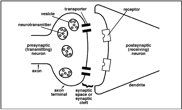
Figure 2.3: The synapse is the site where chemical signals pass between neurons. Neurotransmitter is released from the presynaptic neuron terminals into the extracellular space, the synaptic cleft or synaptic space. The released neurotransmitter molecules can then bind to specific receptors on the postsynaptic neuron membrane to elicit a response.
The brain contains another class of cells called glia. There are as many as ten to fifty times more glial cells than neurons in the central nervous system. Glial cells are categorized as microglia or macroglia. Microglia are phagocytic cells that are mobilized after injury, infection or disease. They are derived from macrophages and are unrelated to other cell types in the nervous system. The three types of macroglia are oligodendrocytes, astrocytes, and Schwann cells. The oligodendrocytes and Schwann cells form the myelin sheaths that insulate axons and enhance conduction of electrical signals along the axons. Scientists know less about the functions of glial cells than they do about the functions of neurons. Glial cells fulfill a variety of functions including:
. Glial cells function as supporting elements in the nervous system to provide structure and to separate and insulate groups of neurons. Oligodendrocytes in the central nervous system and Schwann cells in the peripheral nervous system form myelin, the sheath that wraps around certain axons.
. Some glial cells are scavengers that remove debris after injury or neuronal death.
. Some glial cells buffer the potassium ion (K+) concentration in the extracellular space, and some glial cells take up and remove chemical neurotransmitters from the extracellular space after synaptic transmission.
. Some glial cells guide the migration of neurons and direct the outgrowth of axons during development.
. Some glial cells induce formation of impermeable tight junctions in endothelial cells that line the capillaries and venules of the brain to form the blood-brain barrier.
. Glial cells may serve nutritive functions for nerve cells.
The Blood-Brain Barrier
The blood-brain barrier protects the neurons and glial cells in the brain from substances that could harm the cells. Endothelial cells that form the capillaries and venules make this barrier forming impermeable tight junctions. Astrocytes surround the endothelial cells and induce them to form these junctions. Unlike blood vessels in other parts of the body that are relatively leaky to a variety of molecules, the blood-brain barrier keeps many substances, including toxins, away from the neurons and glia. Blood gases, such as oxygen, and small nutritional molecules do get into the brain.3,4 In addition, drugs of abuse can penetrate the blood-brain barrier. Because most drugs are fat-soluble they can pass through the barrier to reach the brain cells. The blood-brain barrier is important for maintaining the environment of neurons in the brain, but it also presents problems for scientists who are investigating new treatments for brain disorders. If a medication cannot get into the brain to the neurons, it cannot be effective. Researchers attempt to circumvent the problems in different ways. Some techniques attach potential therapeutic agents to molecules that pass through the blood-brain barrier while others attempt to open the blood-brain barrier so that the therapeutic compounds can reach the brain’s neurons.
Neurons Use Electrical and Chemical Signals to Transmit Information
The billions of neurons that make up the brain coordinate thought, behavior, homeostasis, and more. How do all these neurons pass and receive information? Neurons convey information by transmitting messages to other neurons or other types of cells, such as muscles. The following discussion focuses on how one neuron communicates with another neuron. Neurons employ electrical signals to relay information from one part of the neuron to another. The neuron converts the electrical signal to a chemical signal in order to pass the information to another neuron. The target neuron then converts the message back to an electrical impulse to continue the process. Within a single neuron, information is conducted via electrical signaling. When a neuron is stimulated, an electrical impulse, called an action potential, moves along the neuron axon or dendrite.6 Action potentials enable signals to travel very rapidly along the neuron fiber. Action potentials last less than 2 milliseconds (1 millisecond = 0.001 second) and the fastest action potentials can travel the length of a football field in one second. Action potentials result from the flow of ions across the neuronal cell membrane. Neurons, like all cells, maintain a balance of ions inside the cell that differs from the balance outside of the cell. This uneven distribution of ions creates an electrical potential across the cell membrane. This is called the resting membrane potential.
In humans, the resting membrane potential ranges from -40 millivolts (mV) to -80 mV with –65 mV as an average resting membrane potential. The resting membrane potential is, by convention, assigned a negative number because the inside of the neuron is more negatively charged than the outside environment of the neuron. This negative charge results from the unequal distribution of sodium ions (Na+), potassium ions (K+), chloride ions (Cl-), and other organic ions. The resting membrane potential is maintained by an energydependent Na+-K+ pump that keeps Na+ levels low inside the neuron and K+ levels high inside the neuron. In addition, the neuronal membrane is more permeable to K+ than it is to Na+, so that K+ tends to leak out of the cell more readily than Na+ diffuses into the cell.
A stimulus occurring at the end of a nerve fiber starts an electrical change that travels like a wave over the length of the neuron. This electrical change, the action potential, results from a change in the permeability of the neuronal membrane. Sodium ions rush into the neuron, and the inside of the cell becomes more positive. The Na+-K+ pump then restores the balance of sodium and potassium to resting levels. However, the influx of Na+ ions in one area of the neuron fiber starts a similar change in the adjoining segment and the impulse moves from one end of the neuronal fiber to the other. Action potentials are an all-or-none phenomenon. Regardless of the stimuli, the amplitude and duration of an action potential are the same. The action potential either occurs or it doesn’t. The response of the neuron to an action potential depends on how many action potentials it transmits and the time interval between them.
Electrical signals carry information within a single neuron. Communication between neurons (with a few exceptions in mammals) is a chemical process. When the neuron is stimulated, the electrical signal (action potential) travels down the axon to the axon terminals. When the electrical signal reaches the end of the axon, it triggers a series of chemical changes in the neuron. Calcium ions (Ca++) flow into the neuron. The increased Ca++ in the axon terminal then initiates the release of neurotransmitter. A neurotransmitter is a molecule that is released from a neuron to relay information to another cell. Neurotransmitter molecules are stored in membranous sacs called vesicles in the axon terminal. Each vesicle contains thousands of molecules of a neurotransmitter.
For neurons to release their neurotransmitter, the vesicles fuse with the neuronal membrane and then release their contents, the neurotransmitter, via exocytosis. The neurotransmitter molecules are released into the synaptic space and diffuse across the synaptic space to the postsynaptic neuron. A neurotransmitter molecule can then bind to a special receptor on the membrane of the postsynaptic neuron. Receptors are membrane proteins that are able to bind a specific chemical substance, such as a neurotransmitter.
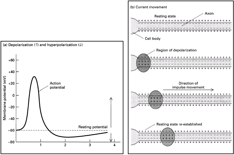
Figure 2.4: (a) Recording of an action potential in an axon following stimulation at time 0 due to changes in the permeability of the cell membrane to sodium and potassium ions. (b) The cell membrane of a resting neuron is more negative on the inside of the cell than on the outside. When the neuron is stimulated, the permeability of the membrane changes allowing Na+ to rush into the cell. This causes the inside of the cell to become more positive. This local change starts a similarchange in the adjoining segment of the neuron’s membrane. In this manner, the electrical impulse moves along the neuron. From: Molecular Cell Biology by Lodish et al. 1986, 1990 by Scientific American Books, Inc. Used with permission by W.H. Freeman and Company.
For example, the dopamine receptor binds the neurotransmitter dopamine, but does not bind other neurotransmitters such as serotonin. The interaction of a receptor and neurotransmitter can be thought of as a lock-and-key for regulating neuronal function. Just as a key fits only a specific lock, a neurotransmitter binds only to a specific receptor. The chemical binding of neurotransmitter and receptor initiates changes in the postsynaptic neuron that may generate an action potential in the postsynaptic neuron. If it does trigger an action potential, the communication process continues. After a neurotransmitter molecule binds to its receptor on the postsynaptic neuron, it comes off of (releases from) the receptor and diffuses back into the synaptic space. The released neurotransmitter, as well as any neurotransmitter that did not bind to a receptor, is either degraded by enzymes in the synaptic cleft or it may be taken back up into the presynaptic axon terminal by active transport through a transporter or reuptake pump. Once the neurotransmitter is back inside the axon terminal, it is either destroyed or repackaged into new vesicles that may be released the next time the neuron is stimulated. Different neurotransmitters are inactivated in different ways.
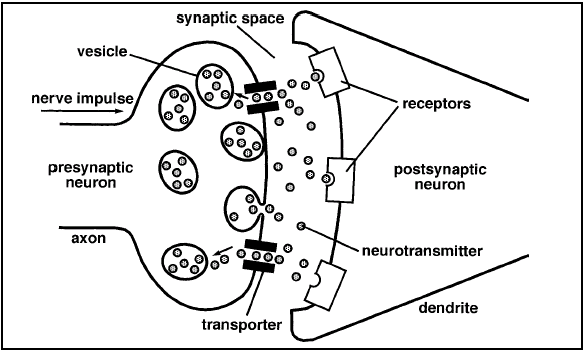
Figure 2.5: Schematic diagram of a synapse. In response to an electrical impulse, neurotransmitter molecules released from the presynaptic axon terminal bind to the specific receptors for that neurotransmitter on the postsynaptic neuron. After binding to the receptor, the neurotransmitter molecules either may be taken back up into the presynaptic neuron through the transporter molecules for repackaging into vesicles or may be degraded by enzymes present in the synaptic cleft.
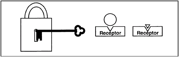
Figure 2.6: Like a lock that will open only if the right key is used, a receptor will bind only a molecule that has the right chemical shape. Molecules that do not have the right "fit" will not bind to the receptor and will not cause a response.
Neurotransmitters Can Be Excitatory or Inhibitory
Different neurotransmitters fulfill different functions in the brain. Some neurotransmitters act to stimulate the firing of a postsynaptic neuron. Neurotransmitters that act this way are called excitatory neurotransmitters because they lead to changes that generate an action potential in the responding neuron. Other neurotransmitters, called inhibitory neurotransmitters, tend to block the changes that cause an action potential to be generated in the responding cell. Table 2.1 lists some of the major neurotransmitters used in the body and their major functions. Each neuron generally synthesizes and releases a single type of neurotransmitter. (Neurons may contain other signaling chemicals, such as neurohormones, in addition to their neurotransmitter.) The postsynaptic neuron often receives both excitatory and inhibitory messages. The response of the postsynaptic cell depends on which message is stronger. Keep in mind that a single neurotransmitter molecule cannot cause an action potential in the responding neuron. An action potential occurs when many neurotransmitter molecules bind to and activate their receptors. Each interaction contributes to the membrane permeability changes that generate the resultant action potential.

Please do the activity below.
This material is based on work supported by the National Institutes of Health under Contract No: 263-98-C-0056. Any opinions, findings, conclusions, or recommendations expressed in this publication are those of the authors and do not necessarily reflect the view of the funding agency.
Copyright c 2000 by BSCS and Videodiscovery, Inc. All rights reserved. You have the permission of BSCS and Videodiscovery, Inc. to reproduce items in this module (including the software) for your classroom use. The copyright on this module, however, does not cover reproduction of these items for any other use. For permissions and other rights under this copyright, please contact BSCS, 5415 Mark Dabling Blvd., Colorado Springs, CO 80918-3842, www.bscs.org, info@bscs.org, (719) 531-5550.
NIH Publication No. 00-4871
ISBN: #1-929614-05-5