| A | B |
|---|
| List the functions of the muscular system | maintain posture, skeletal movement, maintain body temperature, communication, heart beat, respiration, constriction of organs |
| The ability of muscle to shorten forcefully | contractility |
| The capacity of muscle to respond to stimulus | excitability |
| The ability of a muscle to be stretched beyond its normal resting length and is still able to contract | extensibility |
| The ability of muscle to recoil to its original length after it has been stretched | elasticity |
| Name the three major muscle types | skeletal, cardiac, smooth |
| Type of muscle that aids in bone movement | skeletal |
| The type of muscle found in hollow organs. | smooth |
| Type of muscle found only in the heart | cardiac |
| the plasma membrane of a muscle fiber | sacrolemma |
| the threadlike fiber extending from one end of the muscle to the other | myofibril |
| made of Actin and Myosin protein components of muscle fibers | myofilament |
| Highly organized units made of actin and myosin which are joined end to end to form the myofibrils | sarcomere |
| Charge difference across the plasma membrane of an unstimulated muscele cell | resting membrane potential |
| Another term for Electrical Signals | action potential |
| The change in the electrical charge difference across the plasma membrane | depolarization |
| Return of resting membrane potential after the action potential has passed | repolarization |
| A stimulus that is too weak to initiate and action potential | subthreshold stimulus |
| A stimulus that is the minimum stimulus strength require to produce an action potential | threshold stimulus |
| membrane to membrane contact of a nerve cell to a muscle cell | neuromuscular junction |
| Which type of ion channel is Na (sodium) gated and example of? | Ligand-gated |
| When an action potential reaches the presynatic terminal, it causes voltage-gated _________ channels in the plasma membrane of the axon to open and stimulates acetylcholine release | Ca 2+ |
| ________________ binds to receptors of the postsynaptic terminal thereby changing membrane permeability and producing an action potential, which stimulates muscle contraction. | Acetylcholine |
| During muscle contraction what part of the sarcomere does the power stroke cause to move? | myosin |
| The break down of what supplies energy for the recovery stroke of a muscle contraction? | ATP |
| The influx of Ca2+ (calcium) into a muscle fiber causes _____________? | contraction |
| The movement of what ion out of a muscle fiber causes relaxation? | calcium |
| Put the events in one cross-bridge cycle in the correct order | exposure to active site, cross bridge formation, power stroke, cross-bridge release, breakdown of ATP, recovery stroke |
| describe how a muscle contracts | The action potential arrives at the presynaptic membrane, causing Calcium ion voltage gated channels to open. The calcium ions enter the presynaptic terminal. They cause the synaptic vessicles to release the acetylcholine out into the synaptic cleft. The acetylcholine binds to the ligand gated channels on the postsynaptic membrane causing them to open. The sodium ions in the synaptic cleft move into the postsynaptic membrane causing depolarization of the muscle cell and causes a contraction. |
| muscles decreased capacity to work and its reduced efficiency of performance | fatigue |
| What do striations tell you? | direction of pull |
| What happens to the components of the A-band in a sarcomere when a muscle contracts? | actin gets closer to H-zone, myosin doesn't change |
| What do muscle fibers always point to? | attachment points |
| What do muscle fibers show? | direction of pull |
| How do muscles attach to bone? | tendinous (aponeurosis) attachments or fleshy attachments |
| Attachment that pulls on the bone and moves and is usually distal. | insertion |
| The attachment that is immovable and is usually proxinal | origin |
| Muscles that decrease the angle between ventral surfaces. | flexors |
| Muscles that increase the angle between ventral surfaces. | extensors |
| Flexors are always on the ____ side and the extensors are always on the _____ side. | flexors are on ventral side, extensors are on dorsal side |
Label the parts of the actin, 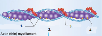 | 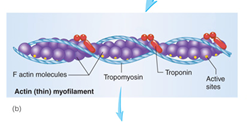 |
Label the parts of the myosin, 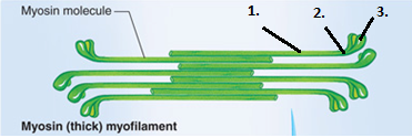 | 1. Rod 2. Hinge region 3. Head |
Label the parts of the sarcomere, 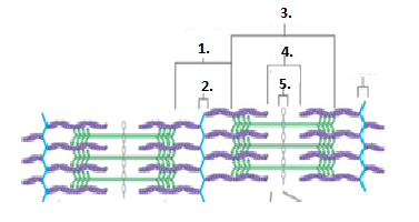 | 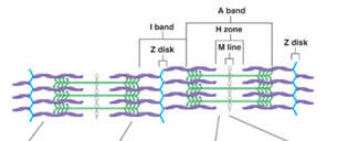 |
Label the parts of the neuromuscular junction,  | 1. presynaptic terminal 2. synaptic vesicles 3. postsynaptic membrane 4. synaptic cleft |
