| A | B |
|---|
| this film is positioned too far occlusally (not far enough up) to include teeth roots | 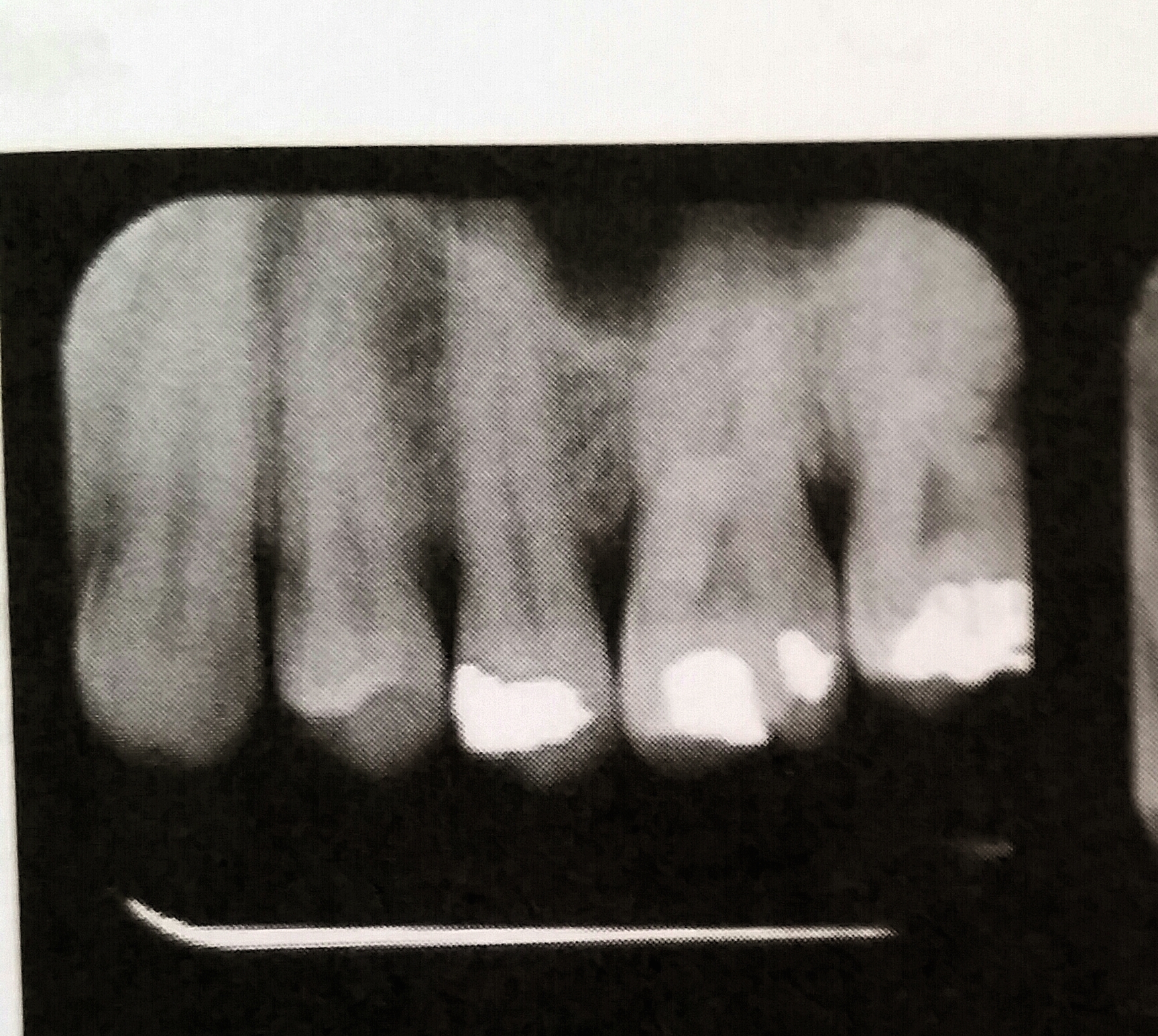 |
the technical error in this radiograph is an example of?,  | elongation, |
Which technical errors are demonstrated in this radiograph,  | film lacement; film must be placed on batwings so equal portions of both maxillary and mandibular crowns of posterior teeth, |
| This helps reduce x-ray exposure for the patient | collimator is a pice of lead that restricts the beam to the size of the film |
| largest intramural film size | #4 used mostly when exposing occlusal radiographs on adults |
| which of the four components of a film packet reduces secondary radiation | lead foil |
| a radiograph that is exposed using a high level of KVp will have | many shades of gray; a radiograph that has low contrast |
| cells with high reproductive rates are considered to be | radiosensitive |
| areas that appear dark on a radiograph are called | radiolucent |
| maximum permissable dose of radiation for an operator | 5 REMS or 0.05 Sv pr year |
| what describes a radiographs darkness | density; describes the opacity of an object to x-rays; denser objects are more opaque |
| overexposure (more than 2-3 minutes) to a safelight will produce a film that is | fogged |
| the purpose of the aluminum filter is to | allow for a more penetrating primary beam; removes low energy, long wavelengths from beam producing more penetrating power |
| which film speed is fastest | F; Fis 20% faster than E and 50% faster than D; A is the slowest |
| what is a major advantage of panoramic radiograph | lower cost to patient compared to intraoral films |
| what structure appears radiopaque on a radiograph | PFM, metal restorations, tooth enamel and dense area of bone |
| what anatomical structures will appear radiolucent | mental foreamen; lacks thickness so is radiolucent |
| which cells are most radiosensitive | bone marrow cells |
| number of waves that pass a given point per unit of time is the | wave frequency |
| if a film is reversed in the mouth during an exposure, resulting image will look | lighter along with herring bone or tire tread effect |
| the period of time between exposure to x-rays and the appearance of radiation damage (biological effects) is | latent period |
| which of the following condition can be identified on a radiograph | salivary stones and root tips; soft tissues is not seen |
| which type of ionizing radiation produce the least amount of scatter radiation | short wavelength radiation |
| how often should automatic processing solutions be changed | every 3-4 weeks |
| elongation of an image on a radiograph may be caused by | insufficient vertical angulation |
| regulations state that an x-ray unit operating above 70 KVp must have a total filtration of | 2.5mm |
| a radiopaque landmark found superimposed over the apical areas of the molars in the maxilla is the | sinus floor |
| when using bisecting angle technique, the vertical angulation of the maxillary premolar exposure is | +30 |
| advantage of using an intensifying screen | reduce exposure time reducing radiation exposure to patient |
| failure to fix film long enough will result in a radiograph with | brown tint |
| negatively charged part of the x-ray tube is the | cathode |
| what is the best way for the assistant to evaluate the amount of radiation exposure | dosimeter at work for 3 months |
| at the patient's skin, the diameter of the x-ray beam should not exceed | 2.75 in |
| excessive vertical angulation causes | foreshortened tooth structures |
| which of the following structures will appear the most radiopaque on a radiograph; amalgam, pulp chamber; paper points; periodontal ligament | pulp chamber |
| if the developer solution is exhausted the radiograph will be | underdeveloped |
| which is a property of an x-ray | no mass or weight |
| maximum permissible yearly dose of radiation that a pregnant women should receive | 50 mSv (10% of what is allowed an occupational worker) |
| What is the minimum distance an operator should stand during an x-ray exposure | 6 ft |
| which of the following would the operator adjust in order to increase the quantity of electrons inside the tube | milliamperage |
| overexposure of the skin to radiation will cause redness or sunburned appearance called | erythema |
| what must be done with nondisposable dental film holder after each use | sterilized |
| the target in the x-ray tube is composed of what material | tungsten |
| identify the anatomical structure that will appear radiolucent on the radiograph | mental foreamen |
| collar or lead apron is used to protect the patient's what? | thyroid gland |
| overlapping images is caused by | incorrect horizontal angulation |
| which of the following is the correct test to detect light leaks in the darkroom | coin test |
| a structure that stops or absorbs x-rays will appear | radiopaque (lighter areas) |
| what type of x-ray produce the radiograph image in the form of a latent image | primary |
| the most penetrating x-rays have a | short wavelength |
| on which x-ray would an inverted "Y" (outline of nasal floor and anterior floor of maxillary sinus) be seen | maxillary canine periapical |
| the RINN XCP is assembled to expose a mandibular right molar periodical; Which area can be exposed without having to reassemble the film holder | maxillary left molar |
| ALARA stands for | as low as reasonably achievable |
| a step wedge is a devise used to test the | automatic processor |
| to avoid cone cuts on a radiograph the operator must | center the PID over the film |
| assuming the mA and the KVp stay the same, if the focal film distance doubles, the exposure time needed to produce a quality fill quadruples; This concept is known as | inverse square law |
| when taking a radiographic survey on a child under the age of 10, the exposure (mAs) should be reduced by | 50% |
| Identify the correct setting that would indicate the PID pointing to the floor | +10; positive angulations indicated PID is pointing toward the floor |
| decreasing the amount of time a duplicating film is exposed to light will result in a film that is | darker; longer exposed to light source the lighter it will be |
| a size #3 film is used for | bitewing radiograph on adult |
| what is responsible for recording of the image on the radiograph | polyester base which contains silver halide crystals |
| anatomic structure present on mandibular anterior radiograph | lingual foreamen |
| bitewing radiographs are most useful in diagnosis of | interproximal decay |
| bitewing radiographs are used to locate | interproximal caries |
| patients are protected from radiation by the filter in the tube head that | eliminates stronger wavelengths |
| when processing radiographs manually the time and the temperature for developing is | 5 minutes at 68 degrees farhenheit |
| film speed is determined by | size of the crystals |
| radiation that impacts future generations is what kind of effect | genetic |
| a properly placed premolar bitewing will include | medial of the canine |
| the mandibular canal is found on which film | mandibular molar PA |
| in which exposure would one identify the genial tubercle as a landmark | mandibular incisor |
| maxillary tuberosity is found on which film | maxillary molar |
| what is not visible in the mandibular molar exposure | mental foreamen |
| Compared to ANSI D-speed film, F-spped film requires less | exposure time |
| old developer solution will cause the films to appear | light |
| what can cause a film to be light | a reversed film |
| the process of removing electrons from atoms is called | ionization |
| what is one factor determining exposure time | size of patient |
| correct horizontal angulation prevents which of the following radiographic areas | foreshortening |
| what is the purpose of a risk management program for taking radiographs in a dental office | reduce chance of malpractice suit |
| an x-ray machine operating at 70kVp must have how much aluminum filtration in order to comply with federal law | 2.5mm |
| static from improperly handling film will appear to have which pattern on the finished film | lightening |
| the glass wall and insulating oil acts as what type of filtration for photons generated in the tube head | inherent |
| the total amount of dental radiation a patient receives | is cumulative over a lifetime |
| example of periodical radiograph | 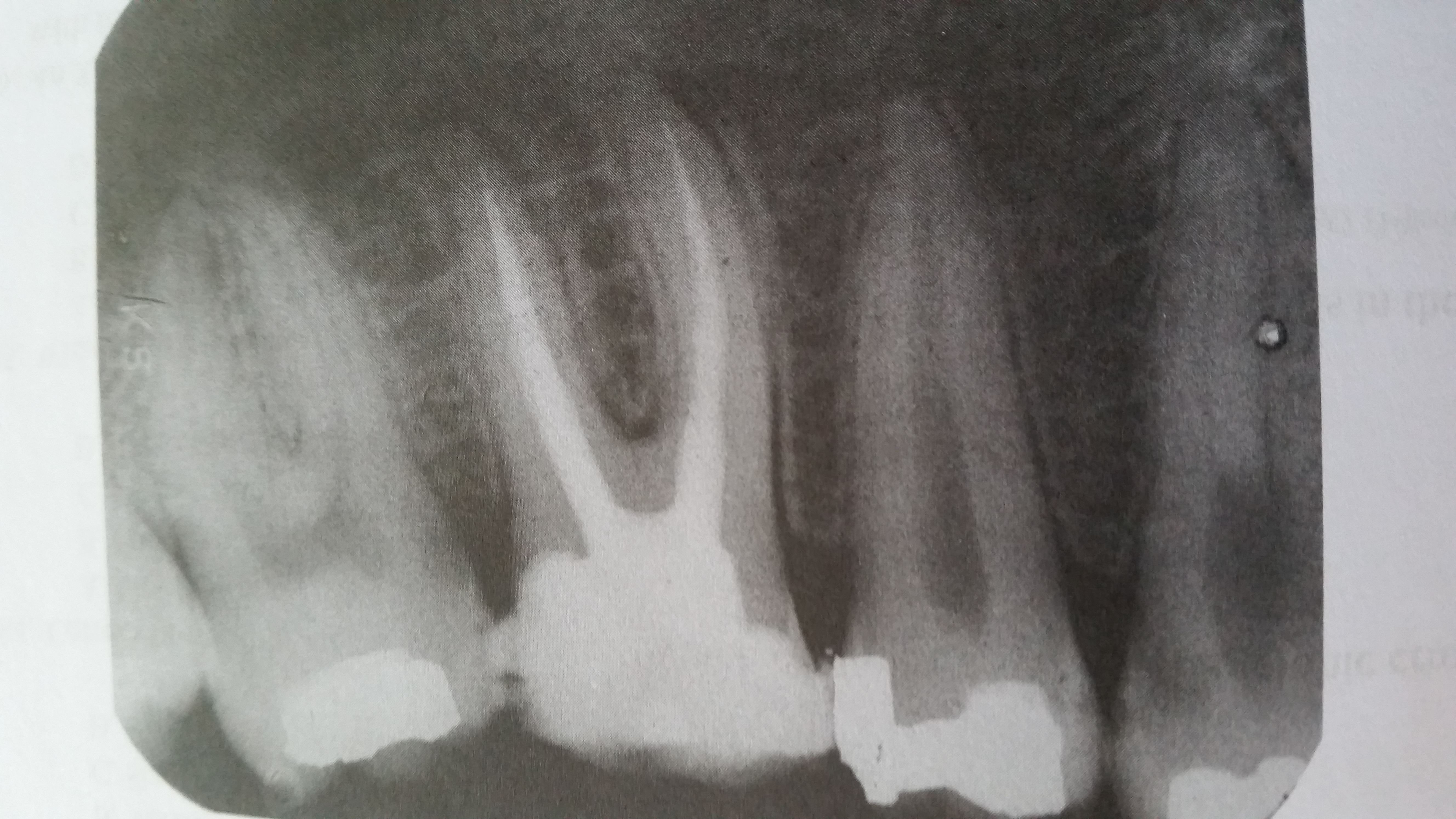 |
| what size film is used for periodical exposure of posterior teeth | Size 2 |
| rapaque areas visible in the medial and distal roots of molars are, | root canals with gutta percha |
| what is the restoration visible in second molar is a, | DO amalgam |
What tooth is missing, 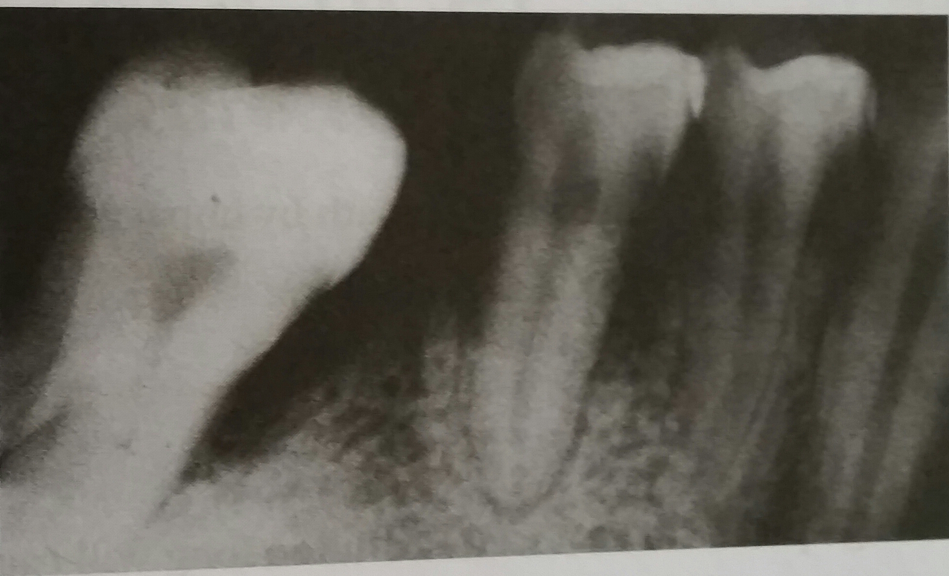 | #30 |
| the radiolucent area involving molars and premolars shows, | severe bone loss |
| which of the following is the earliest clinical sign of overexposure to x-rays | erythema |
| which of the following is considered to be one the yearly permissible dose for dental patient | 10mSv/0.1 REM |
| the primary purpose of monitoring X-ray equipment is to | check for radiation leaks |
| primary advantage of automatic film processor | no need for chemicals |
| during x-ray exposure a thyroid collar is placed on | all patients |
| radiation that has been deflected from its path by impact with matter is called | scatter radiation |
| leaving the radiograph in the final water bath too long will cause the image to | become clear |
this radiograph is an example of, 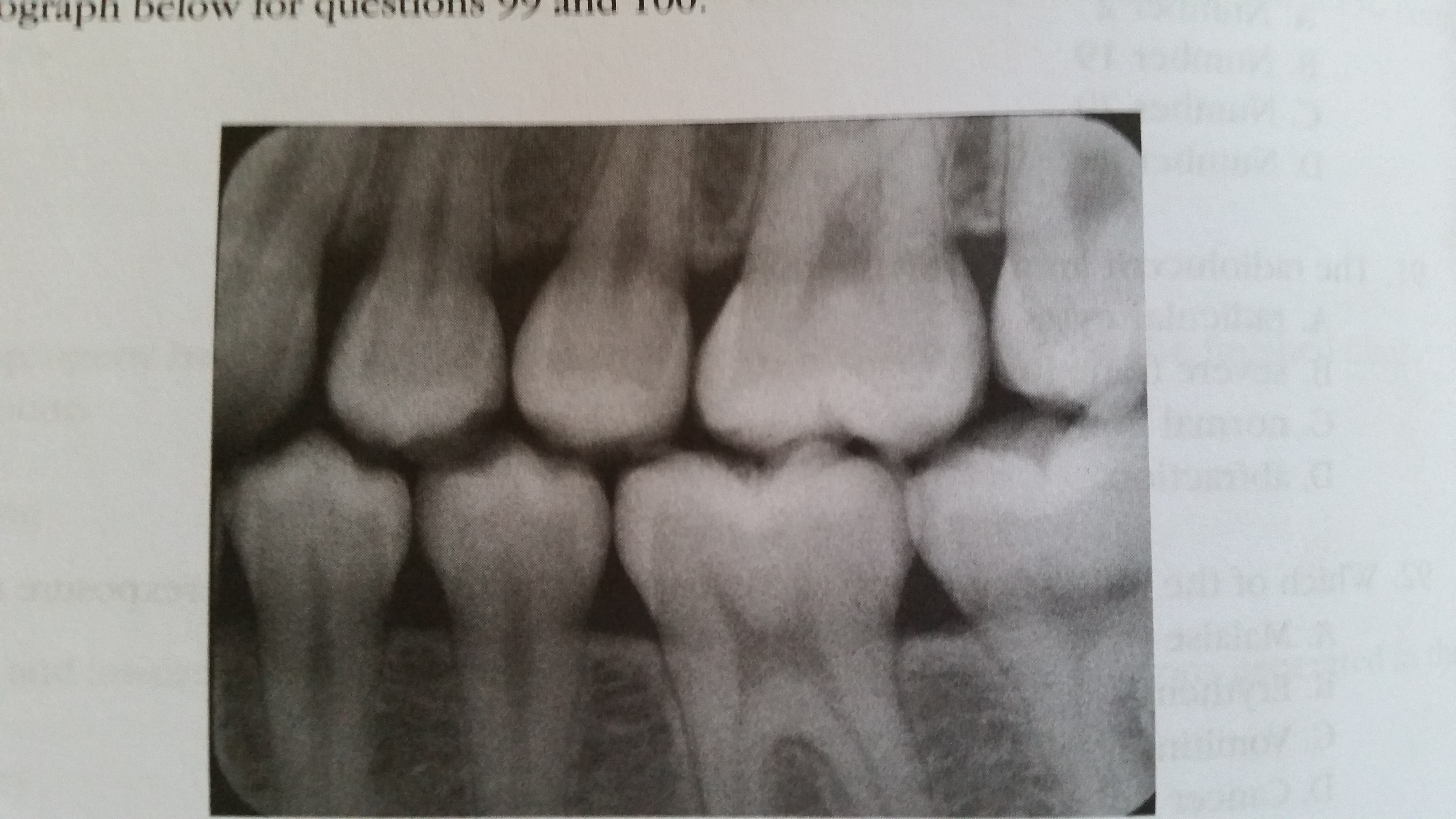 | bitewing |
| this radiograph illustrates the, | left premolar |
